
Overview
This intensive, hands-on workshop is designed for nurse anesthetists seeking to enhance their skills in intravenous (IV) cannulation using ultrasound guidance. The course integrates foundational knowledge with practical application, focusing on improving success rates, patient comfort, and procedural safety in challenging vascular access scenarios.
Learning Objectives
Participants will:
- Understand the principles of ultrasound physics and machine operation (“knobology”).
- Gain insight into upper extremity vascular anatomy relevant to IV access.
- Learn to select appropriate catheters based on vein characteristics and clinical context.
- Develop and refine hand-eye coordination for real-time ultrasound-guided cannulation.
- Practice ultrasound-guided cannulation techniques in a controlled, supportive environment.
Workshop Content
1. Ultrasound Physics and Knobology
The workshop begins with a concise theoretical session covering the basics of ultrasound physics, including sound wave propagation, image generation, and artifact recognition. Participants will also learn the practical aspects of operating an ultrasound machine—adjusting depth, gain, probe orientation, and optimizing image quality for vascular access.
2. Vascular Anatomy
A focused review of the vascular anatomy of the upper extremity will be provided, emphasizing the identification of superficial and deep veins, anatomical landmarks, and common anatomical variations. This knowledge is essential for safe and effective cannulation.
3. Catheter Selection
Participants will be guided through the principles of catheter selection, including gauge, length, and material, based on vein depth, diameter, and clinical indication. Emphasis will be placed on matching catheter characteristics to patient-specific vascular anatomy and procedural goals.
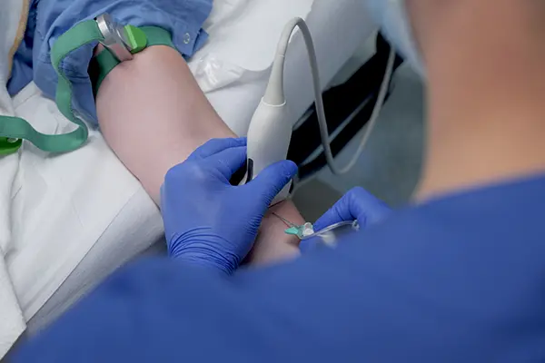
4. Hands-On Training (Core Component)
The hands-on portion is the heart of the workshop and is divided into two progressive phases:
Part 1: Anatomy Identification on a Live Model
Participants will work in small groups to trace and identify veins on a live model (a colleague-trainee), without performing cannulation. This session allows trainees to:
- Develop probe-hand coordination.
- Appreciate vein compressibility, depth, and diameter.
- Practice dynamic scanning techniques in both transverse and longitudinal planes.
- Build confidence in identifying target veins and surrounding structures.
Part 2: 1:1 Cannulation Practice on a Non-Human Tissue Model
In this individualized session, each participant will perform ultrasound-guided IV cannulation on a high-fidelity tissue model. The focus will be on:
- Visualizing the needle tip in real-time.
- Deforming the target vein to confirm contact.
- Maintaining eye-hand coordination while manipulating the probe and inserting the catheter.
- Confirming successful cannulation under ultrasound visualization.
This phase emphasizes the critical skill of maintaining continuous visualization of the needle tip to avoid complications and ensure accurate placement.
Why This Workshop Matters
Ultrasound-guided IV access is a vital skill in modern clinical practice, especially in patients with difficult venous access. Mastery of this technique reduces the number of attempts, improves patient satisfaction, and enhances procedural safety. Training in ultrasound physics and hands-on practice are essential prerequisites for competent and confident application in clinical settings.